研究目的
Investigating the formation, sizes, and degree of crystallinity of silicon nanoparticles (SNPs) produced via picosecond laser ablation of porous silicon and silicon microparticles in water, and their potential applications in photonics and biomedicine.
研究成果
The study successfully demonstrated the formation of SNPs with sizes ranging from 50 to 300 nm via laser ablation and fragmentation, showing high crystallinity, especially in the case of laser fragmentation of micropowders. These findings suggest promising applications in photonics and biomedicine, with potential for further research to optimize nanoparticle properties for specific uses.
研究不足
The study focuses on the structural analysis of SNPs formed via laser ablation and fragmentation, with limited discussion on the optimization of the process for specific applications. The potential for further refinement of nanoparticle sizes and crystallinity for enhanced photonic and biomedical applications is noted.
1:Experimental Design and Method Selection:
The study involved picosecond laser ablation of porous silicon and silicon microparticles in water to form SNPs. Raman spectroscopy was used to analyze the crystallinity of the nanoparticles.
2:Sample Selection and Data Sources:
Micro- and mesoporous silicon layers were prepared from boron-doped single-layer silicon wafers. A commercial micropowder of chemically pure silicon was used for laser fragmentation.
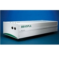
3:List of Experimental Equipment and Materials:
A Nd:YAG EKSPLA PL 2143A picosecond laser, Carl Zeiss Supra 40 electron microscope for SEM, and Horiba Jobin Yvon HR 800 spectrometer for Raman spectroscopy.
4:Experimental Procedures and Operational Workflow:
Laser ablation was performed on PS layers and micropowder suspensions in distilled water. The exposure time was 30 min. Post-irradiation, SNP suspensions were analyzed via SEM and Raman spectroscopy.
5:Data Analysis Methods:
The degree of crystallinity was evaluated using Raman line intensities for crystalline and amorphous phases, with the volume fraction of the amorphous phase calculated from the integral intensities of these peaks.
独家科研数据包���,助您复现前沿成果,加速创新突破
获取完整内容